Theory of Light and Color

18. The Human Eye and the Camera Eye (Part 1)
Both the human eye and the camera eye are essentially imaging devices with the same basic structure. However, if we look at the details, there are many differences.
Imaging Optics (Imaging Lens and Cornea/Lens)
Both have an optical system with a similar configuration. The convex lens of the human eye is composed of the cornea and a crystalline lens. Cameras have multiple lenses, which function as a convex lens. In both cases, the lens forms an inverted image of the subject on the imaging surface.
Cameras and the human eye have different ways of adjusting focus. A camera is designed to adjust its focus by moving the lens back and forth along an optical axis. On the other hand, a human eye's crystalline lens changes its thickness to adjust the refractive index of the convex lens. The center of the crystalline lens becomes thicker at shorter distances and thinner when looking far away.
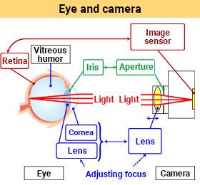
Image Plane (Imaging Sensor and Retina)
In a camera, an image sensor is on a flat surface. The sensitivity and resolution of the image sensor are uniform across the entire imaging surface.
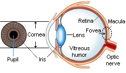
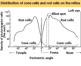
The image plane in the human eye is the retina, which spreads over the spherical fundus. There are various photoreceptor cells (cone cells and rod cells) on the retina, but the density of their distribution is not uniform. Cone cells are concentrated in the center of the retina (fovea centralis) and only slightly distributed in other parts. In contrast, rod cells are located not in the fovea centralis but in other parts of the fundus.
Sensitivity and resolution are high in the area surrounding the fovea where the cone cells are concentrated. When we read a book or do detailed work, we focus our gaze on that area. The eye unconsciously focuses on the image formed around the fovea centralis, which has the highest imaging performance. For instance, letters at the edge of the field of view are difficult to read. These letters are far from the fovea centralis on the retina, in an area where the concentration of cone cells is low. Therefore, you cannot perceive it clearly as a high-resolution image even though you know it exists.
The fovea centralis and its surroundings look like a yellow spot, and this pigmented area is called the macula. Since cone cells are instrumental to visual acuity and color vision, it is thought that the yellow macula protects them from incoming light.
As mentioned in Chapter 2, the shorter the wavelength of light, the greater the physical energy of the photon, which increases stress on the cone cells. Yellow has low transmittance of short wavelengths and high transmittance of mid and long wavelengths in the visual spectrum. So the macula tissue covering the cone cells functions as a filter that protects the cone cells. Thus, the spectral response of cone cells is different in the macula compared to other parts of the eye.
In general, human color vision perceives slightly different colors depending on the viewing angle, which is called the area effect. For this reason, there are two standards for color matching functions to evaluate colors: 2° field of view and 10° field of view. The 2° field of view is mainly focused on the macula, whereas the 10° field of view includes the wider area outside the macula. Thus, the color-matching function of the 2° field of view has slightly longer wavelengths than that of the 10° field of view. ≪1≫
Blind Spot
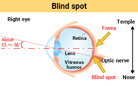
There is an area on the retina where no photoreceptor cells exist. Optic nerve fibers from photoreceptor cells distributed throughout the retina are bundled here and connect to the brain. It also serves as the entrance and exit point for blood vessels that supply blood to various tissues on the retina. Because there are no photoreceptor cells, the retina cannot receive any images formed in this spot, like a hole in the field of vision.
This blind spot occurs about 15 to 16 degrees from the optical axis that connects the centers of the imaging lens (cornea and crystalline lens) and the fovea centralis. It is almost unnoticeable, as the brain fills in the blind spot by estimating information from the surrounding areas. This function is unique to the human eye, but digital cameras have a similar feature. ≪2≫
Light Adaptation in the Human Eye vs. Camera Exposure
Three basic factors determine camera exposure: aperture value (f-number), exposure time (shutter speed), and image sensor sensitivity (ISO sensitivity). The quantity of light (time-integral value of illuminance on the imaging surface) is adjusted so that the maximum and minimum luminance is within the sensor's dynamic range.
For example, if the aperture range is from F2 to F64 (10 aperture steps), the adjustable exposure range is 210. ≪3≫ Another way to control exposure is through shutter speed. Exposure time can range from high speed (ex. 1/4000 sec) to low speed (ex. 30 sec). If a camera has 17 steps of shutter speed, its adjustable range is 217. With such specifications, the camera's exposure can be adjusted within a range of 210 X 217 = 227 ≒ 108.
The standard sensitivity of an image sensor is ISO 100. Depending on the subject and goal of the image, the sensitivity may be higher, such as ISO 400 and ISO 800, or lower, such as ISO 50 and ISO 25. This corresponds to the (physiological) change in the sensitivity of photoreceptor cells.
The human eye's equivalent of the camera aperture is the iris and pupil. The size of the pupil is about 2 mm in diameter in bright environments and changes to about 8 mm in the dark. The adjustable range of exposure of the iris is only 4 times the pupil diameter ratio and thus 16 times its area ratio. Therefore, most of the eye's exposure control is a physiological response to changes in photoreceptor sensitivity. The cone cells are sensitive to changes in brightness. However, they stop working when brightness levels become too low. Rod cells become active instead, though it takes them several minutes to adjust to their highest level of sensitivity.
| Function | Eye | Camera | ||||
|---|---|---|---|---|---|---|
| Blocking incident light | Eyelid | Shutter & lens cap |
||||
| Imaging & focus adjustment |
Cornea & crystalline lens |
The cornea has a refractive power of 3/4 to 2/3 of the entire eyeball. Changing the crystalline lens thickness adjusts the refractive power of 1/4 to 1/3 of the eye. (Auto-focus) |
Imaging lens | Adjusting the lens position back and forth on the optical axis. | ||
| Light sensor | Retina | It is arranged spherically on the fundus. It has a sensitivity distribution and a resolution sensitivity. The cone cells are concentrated in the macula and fovea centralis. The rod cells are widely distributed outside the fovea centralis. |
Image sensor | Fixed to a flat imaging surface. Uniform sensitivity and resolution. |
||
| Light sensor adjustment (exposure) |
Iris & retina sensitivity |
Iris diameter: 2 to 8 mm (Area ratio = 1:16) Working illuminance range of the visual system: 103 ~ 105 (Auto exposure) |
Aperture & exposure time |
Ex. Aperture for F2 ~ F64: 210 ≒ 103 times Exposure time for 1/4000 ~ 30 seconds: 217 ≒ 105 times Aperture x Exposure time: 103 x 105 = 108 times |
||
Comment
≪1≫ 2° field of view and 10° field of view
JIS Z 8701 (1999)
≪2≫ Defective pixel correction
Digital cameras are equipped with CCD or CMOS image sensors. These image sensors are composed of over 10 million fine pixels. Various factors in production can cause pixels to malfunction (defective pixels).
Excluding cases where there are too many defective pixels, the output signal of the normal pixels around the defective pixel can be averaged and treated as the signal of the defective pixel. This process is similar to how the human eye corrects its blind spot.
≪3≫ Relationship between shutter speed and exposure
The exposure time (shutter speed) T and aperture value (f-number), which control the camera's exposure, are shown in the table below. The f-number is defined according to the aperture diameter. Since the luminous flux passing through the aperture is proportional to its area, the exposure is proportional to the square of the f-number. If you increase the f-number by n, the exposure decreases by 1/2n, and if you decrease by n, the exposure increases by 2n.
| The exposure time (shutter speed) T | ・・・・・, 1/4000, 1/2000, 1/1000, 1/500, 1/250, 1/125, 1/60, 1/30, 1/15, 1/8, 1/4, 1/2, 1, 2, 4, ・・・・・ |
|---|---|
| Aperture value (F-number) | 1, 1.4, 2, 2.8, 4, 5.6, 8, 11, 15, 22, 30, 45, 60, 90, ・・・・・ |